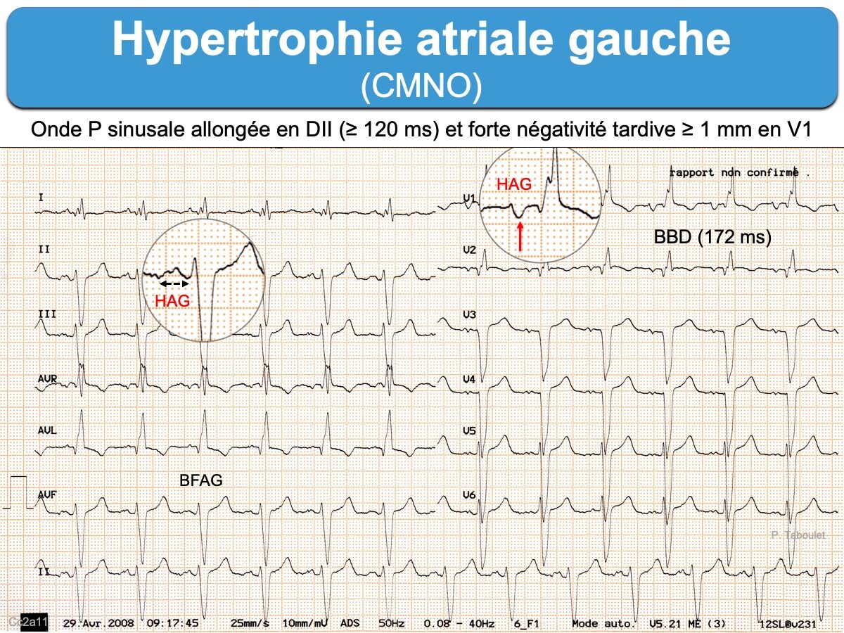
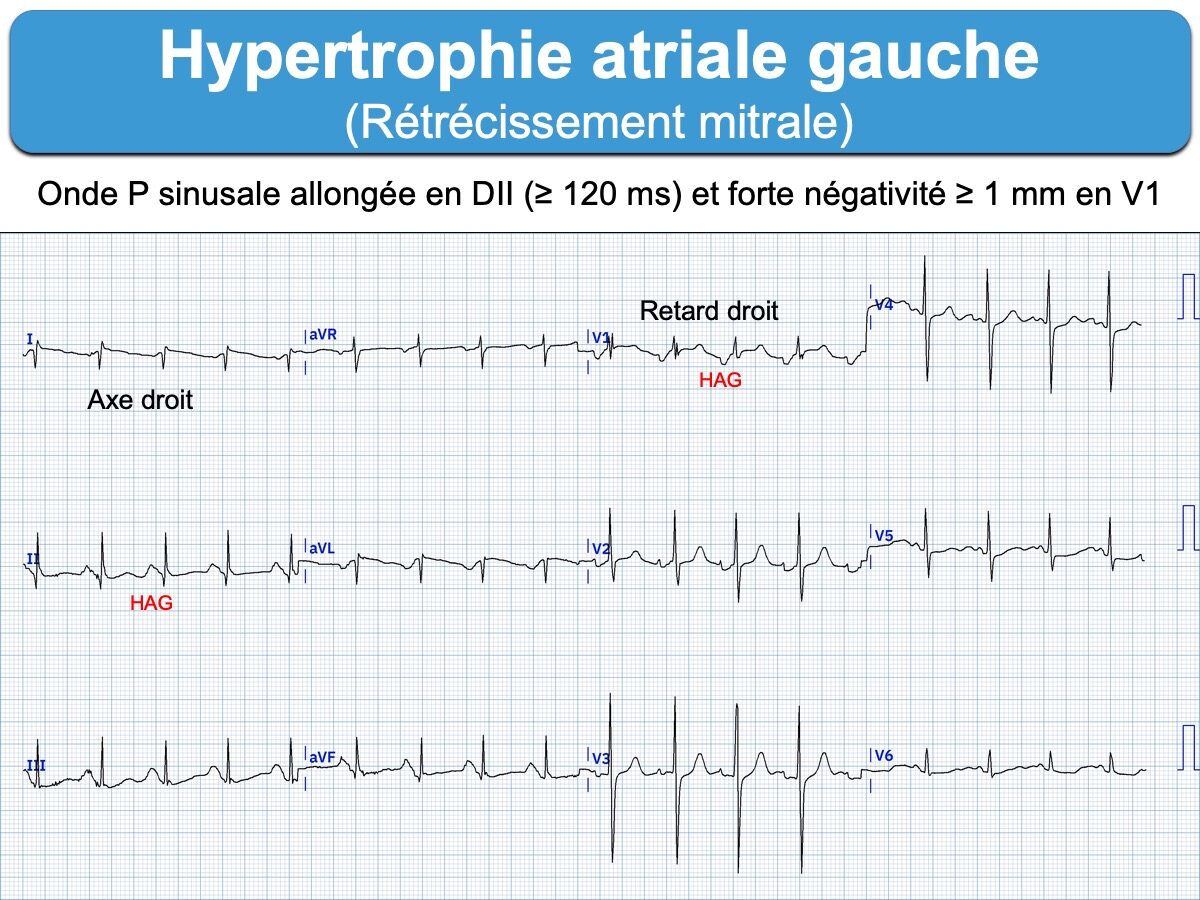
Une hypertrophie/dilatation de l’oreillette gauche (left atrial enlargement) peut prolonger la durée et modifier la morphologie deux derniers tiers de la dépolarisation atriale (cf. Onde P sinusale).
Définition ECG
Une HAG électrique est définie par :
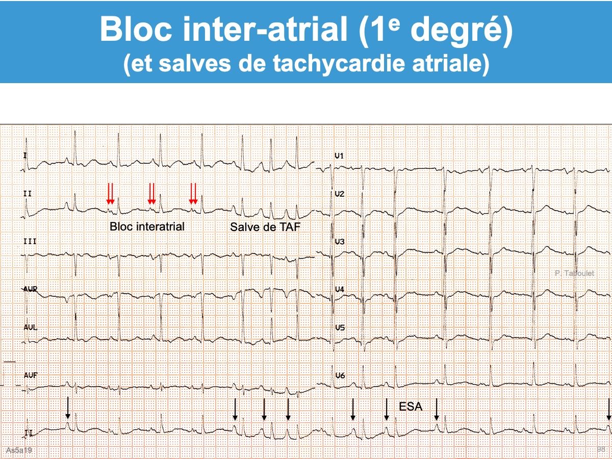
- En DII. Une onde P sinusale allongée dont la durée est ≥ 120 ms (3 petits carreaux horizontaux ou 3 mm en étalonnage 10 mm = 1 mV). L’onde PDII est souvent bifide avec une deuxième double bosse ≥ 40 ms (AHA 2009 [1]). Néanmoins, une anomalie de conduction intra-atriale peut donner une prolongation similaire de l’onde P (Cf. Bloc inter-atrial) [1][3]. Pour cette raison, le terme de left atrial abnormality est préféré au terme hypertrophie/dilatation par les auteurs américains [1][3]. D’autres anomalies de l’onde P ont été rapportées comme une déviation axiale gauche de P de 15° vers la gauche ou de la portion/force terminale de P [3].
- En V1. Une onde P à forte polarité terminale négative est aussi évocatrice d’une HAG (amplitude ≥ 1 mm ou mieux surface de P- > P+ ou double produit de l’amplitude par la durée de l’onde P terminale) [3]. Cette anomalie traduit un important remodelage atrial et augmente le risque de « stroke » (AVC [4]).
- L’HAG électrique présente une bonne spécificité (mais une sensibilité mauvaise) pour le diagnostic d’HAG anatomique, comparée avec les données de l’IRM [2].
La sensibilité de l’ECG pour le diagnostic d’une HAG est mauvaise, comparée aux données de l’échocardiographie ou de l’IRM. Néanmoins, l’existence d’une HAG électrique au côté d’une anomalie des complexes QRS augmente la probabilité de cardiopathie aiguë ou chronique avec hypertrophie ventriculaire gauche.
Etiologies
Une HAG se rencontre de façon typique au cours de l’évolution de la plupart des cardiopathies gauches (hypertension artérielle, cardiomyopathie, rétrécissement aortique, valvulopathie mitrale…).
Diagnostic différentiel
Le bloc inter-atrial qui correspond à une anomalie de conduction au sein de l’oreillette gauche ou inter atrial. Néanmoins, les deux anomalies sont souvent associées et difficiles à séparer l’une de l’autre. Le terme
[1] Hancock EW et al. AHA/ACCF/HRS recommendations for the standardization and interpretation of the electrocardiogram: part V: electrocardiogram changes associated with cardiac chamber hypertrophy... J Am Coll Cardiol. 2009;53(11):992-1002. Review. (téléchargeable)
–> Left atrial abnormality usually involves prolongation of the total atrial activation time, because left atrial activation begins and ends later than right atrial activation. Delay in the left atrial activation tends to cause a double-peaked or notched P wave, because the right and left atrial peaks that are normally nearly simultaneous and fused into a single peak become more widely separated. Activation of the left atrium has a more leftward and posterior vector than that of the right atrium. The product of the amplitude and the duration of the terminal negative component of the P wave in lead V1 (the P terminal force) has been used most frequently of the various criteria for left atrial abnormality, but the P-wave duration (120 ms or more) and widely notched P wave (40 ms or more) appear to have equal value. Several other criteria, including left axis of the terminal P wave ( 30 to 90), and possibly the P-wave area, are also useful.59,60 A purely negative P wave in V1 is suggestive but can occur without an increased P terminal force.
[2] Tsao CW, Josephson ME, Hauser TH, et al. Accuracy of electrocardiographic criteria for atrial enlargement: validation with cardiovascular magnetic resonance. J Cardiovasc Magn Reson. 2008 Jan 25;10(1):7. (téléchargeable)
The prevalence of CMR LAE and RAE was 28% and 11%, respectively, and by any ECG criteria was 82% and 5%, respectively.
Though nonspecific, the presence of at least one ECG criteria for LAE was 90% sensitive for CMR LAE. The individual criteria P mitrale, P wave axis < 30 degrees , and negative P terminal force in V1 (NPTF-V1) > 0.04s.mm were 88-99% specific although not sensitive for CMR LAE.
ECG was insensitive but 96-100% specific for CMR RAE.
Conclusion: The presence of at least one ECG criteria for LAE is sensitive but not specific for anatomic LAE. Individual criteria for LAE, including P mitrale, P wave axis < 30 degrees , or NPTF-V1 > 0.04s.mm are highly specific, though not sensitive. ECG is highly specific but insensitive for RAE. Individual ECG P wave changes do not reliably both detect and predict anatomic atrial enlargement.
[3] Surawicz B et Knilans TK. Chou’s electrocardiography in clinical practice. Ed. Elsevier, 6th édition 2008. Atrial depolarization (page 37)
Abnormal P waves should usually be referred to as right or left “atrial abnormality” rather than enlargement, overload, strain, or hypertrophy.
[4] Li M, Ji Y, Shen Y, Wang W, Lakshminarayan K, Soliman EZ, Chen M, Chen LY. Deep terminal negative of the P wave in V1 and incidence of ischemic stroke: The atherosclerosis risk in communities (ARIC) study. J Electrocardiol. 2024 Apr 16;84:123-128. doi: 10.1016/j.jelectrocard.2024.03.016. Epub ahead of print. PMID: 38636124.


